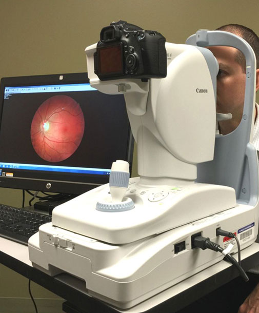
Fundus Photography
Advanced Retinal Imaging
Fundus photography is a powerful diagnostic tool employed at Divyajyoti Eye Hospital to provide a comprehensive view of the back of your eye, including the retina and optic nerve.
This advanced imaging technique allows us to assess the health of these critical structures and detect various eye conditions with exceptional precision, ensuring timely intervention and treatment.
Key Benefits of Fundus Photography
- Early Detection: Enables detection of eye diseases such as diabetic retinopathy, age-related macular degeneration (AMD), and glaucoma before significant vision loss occurs.
- Detailed Evaluation: Provides high-resolution images to examine the retina’s intricate layers and the optic nerve head for any abnormalities.
- Treatment Planning: Guides our ophthalmologists in developing personalized treatment plans tailored to your specific condition.
- Progress Monitoring: Allows us to compare images over time to monitor disease progression and the effectiveness of treatments like laser therapy or injections.
- Patient Education: Provides a visual representation of your eye health, helping you understand your condition and the importance of treatment compliance.
Common Questions about Fundus Photography
Your pupils will be dilated with eye drops. You will then sit in front of a specialized camera, rest your chin on a support, and look at a fixation light while the photographer takes flash pictures of your eye.
The photography itself takes only a few minutes. However, pupil dilation requires about 15-30 minutes to take effect before the photos can be taken.
Because your eyes will be dilated, your vision will be blurry, and you will be sensitive to light for a few hours. It is recommended to bring a pair of sunglasses and have someone else drive you home.